Dry eyes can occur when there is too little aqueous (salt water) tear production and/or too rapid tear evaporation. When the lacrimal gland fails to produce enough tears, the surface of your eye dries out and often causes burning, stinging, and a sandy or gritty sensation. Artificial tears and lubricating ointments may help relieve dry eye symptoms. If these lubricants do not help, closing the tear drain may be helpful. If the lacrimal gland is working properly, but your tears do not drain through the tear duct because it is blocked, your eye may feel watery and tears may even overflow onto your cheek. In some cases, you may be able to simply apply warm compresses and use antibiotic drops. Rapid tear evaporation is believed to be the result of a chronic, low-grade inflammation of the eyelids and particular oil-producing glands in the lids – the most likely culprit in the majority of dry eye cases. Frantz EyeCare is one of only a handful of practices in Florida to intensively focus on this important issue and devote the personnel, resources, and wide array of advanced treatments to offer effective management of this problem to our patients.
When managing a patient’s dry eye complaint, we must first identify the type of dry eye you are experiencing (meibomian gland dysfunction, aqueous deficiency, allergy-related, contact lens-related, or blepharitis related). We utilize several tools necessary to properly diagnose and distinguish between the types of dry eye.
Tear Osmolarity Test
Frantz EyeCare is certified by TearLab as an “Accredited Dry Eye Center”. Certification indicates that Frantz EyeCare has demonstrated expertise and dedication to the most advanced technologies and treatments in the management of dry eye disease. Our team of doctors have extensive experience in the diagnosis and treatment of dry eye disease. This test is the latest technology in diagnosing and analyzing the Osmolarity (salt content) of your tears. With the TearLab Osmolarity test, our doctors can measure and assess the health and stability of your tear film. Treatment is prescribed based on the measurements from the TearLab. Treatments range from Omega 3 supplements, tear replacement drops, punctal plugs, anti-inflammatory drops, oral antibiotics and prescription strength artificial tears.
Some of the symptoms of dry eye disease include:
Redness
Burning
Itching
Fluctuation in vision
Feeling of sand or grit in the eye
Contact Lens discomfort
Light Sensitivity
Watery eyes
Tired eyes
Causes of Dry Eye
The lacrimal glands, located behind the upper eyelids, produce tears that moisten our eyes. When we blink, the eyelids spread a film of fresh tears across the eye and pump excess tears into the nasolacrimal duct or tear drain. Without this mechanism working correctly, dry eyes become a chronic problem.
When the lacrimal gland fails to produce enough tears, the surface of your eye dries out and often causes burning, stinging, and a sandy or gritty sensation. Artificial tears and lubricating ointments may help relieve dry eye symptoms. If these lubricants do not help, closing the tear drain may be helpful or in extreme cases surgery may be required to protect your eye.
If the lacrimal gland is working properly, but your tears do not drain through the tear duct because it is blocked, your eye may feel watery and tears may even overflow onto your cheek. In some cases, you may be able to simply apply warm compresses and use antibiotic drops; but often, surgery to relieve the obstruction is the most effective treatment.
Meibomian Gland Disorder (MGD)
Lipid layer testing and blink assessment. The LipiView system evaluates your blink rate and measures lipid layer thickness. The lipid layer is important to keep the ocular surface smooth and comfortable. Patients who do not blink completely are often surprised by test results that demonstrate the incomplete blink. Measurements from the system give us objective “hard evidence” of meibomian gland dysfunction (MGD) and further allow us to see if treatment is improving the condition of your ocular surface. The ocular surface relies on the oil in the tears to keep it wet and smooth for the blink. As the meibomian glands clog, they can become damaged. The longer they are clogged, the more damage occurs and eventually the gland structure can become atrophied.
MGD is a chronic abnormality of the meibomian glands, commonly characterized by terminal duct obstruction and/or changes in the glandular secretion. Patients with MGD can benefit from mechanical expression of blocked meibomian glands using the innovative LipiFlow Thermal Pulsation System. LipiFlow is the only FDA-cleared device for removing gland blockages and restoring gland function. The treatment utilizes heat applied to the inner eyelids and massage to remove the obstructions in your meibomian glands. Research shows significant improvement in both the number of meibomian glands yielding clear liquid and patient-reported dry eye symptoms among patients treated with LipiFlow.
Blepharitis Treatment
Blepharitis, a chronic inflammatory disease of the eyelids, is evident in many patients who have dry eye. We can use an in-office procedure called BlephEx that exfoliates the eyelids while removing scurf and debris. With BlephEx a mechanical device scrubs the lids at the lash line, a deeper cleaning than you can do at home.
Lacrimal Disorders
Blockage within the lacrimal drainage system can keep tears from draining into the nose, causing the tears to build up on the lower eyelid and spill over onto the face. The nasolacrimal duct, a bony canal carrying tears into the nose, is the most common site of obstruction.
The main symptom is constant tearing from one or both eyes with tears running down the face. Because access into the nose is blocked, mucous builds up in the lacrimal sac making the patient prone to infection. Infection in the lacrimal sac can be serious as it can spread to the face, orbit, and brain. This condition is treated initially with antibiotics followed by surgical correction of the obstructed duct.
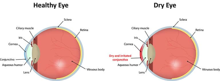
The conjunctiva is the clear membrane that encircles and protects the eyeball. When you look at the white of the eye, you are really looking through the conjunctiva at the sclera – which is the tough, leathery outer coat of the eye. The conjunctiva has many small blood vessels running through it. The purpose of the conjunctiva is to lubricate and protect the eye and to allow it to move in its socket.
Conjunctivitis is an inflammation of the lining of the eye. It can be caused by bacteria (as in “pink eye”), viruses, chemicals, allergies, and more. It is sometimes difficult to tell exactly which is the real cause.
Bacterial conjunctivitis is characterized by swelling of the lid, a yellowish discharge, sometimes a scratchy feeling in the eye (often both eyes), and itching and mattering of the lids, especially in the mornings upon awakening. The conjunctiva is red and sometimes thickened. The bacteria most commonly at fault are staphylococcus, streptococcus, and H. influenza. This disease is very contagious, and sometimes entire families can become infected. Laboratory cultures are not typically used to make the diagnosis since this is expensive and time-consuming. Most infections are over by the time the results of the lab tests come back.
Treatment is curative. Usually, antibiotic drops and compresses ease the discomfort and clear up the infection in just a few days. Occasionally, the infection does not respond well to the drops. In those rare cases, a second visit to the office should be made and other measures undertaken. In severe infections, oral antibiotics are necessary. Covering the eye is not a good idea because this incubates the germs.
If left untreated, conjunctivitis can create serious complications, such as infections in the cornea, lids, and tear ducts. Prevention is important for avoiding the disease and stopping its spread. Careful washing of hands, and the avoidance of touching your eyes or sharing things with contagious individuals are all helpful. Little children frequently get conjunctivitis because of their lack of understanding about hygiene and the resulting contact with germs.
Along the upper and lower lids are located a number of glands that manufacture part of the tear film that protects and lubricates the eyeball. If one of these glands becomes blocked, a small lump forms. This is called a chalazion (chalazia, plural).
Chalazia may vary in size from small, almost invisible lumps to rather large masses as big as a little fingernail. Sometimes tender in their early stages, they are later painless and frequently will form a firm swelling in the lid. This lump can distort the eyeball, causing blurred vision if left untreated. Chalazia are not caused by infection; however, they may become a site for infection once they have become established. Their exact cause remains unknown; however, there are several conditions associated with chalazia such as: seborrhea, chronic lid inflammation, dry eyes, and acne. Most chalazia will disappear in a few weeks without any special therapy. To help them go away, frequent hot packs throughout the day and drops are helpful, especially in the early stages. In some cases, oral medications can help prevent recurrences. If a chalazion persists, a simple in-office surgical procedure can be performed to remove it. The chalazion is drained from the inside of the lid after a small injection of a local anesthetic. There is no visible scar and healing is rapid and painless. Often the eye is patched overnight to ensure proper healing.
If you think you are suffering from the above symptoms, be sure to mention any dry eye concerns at your next visit so you can experience the Dry Eye Center of Excellence.